The wrist is the most commonly injured part of the arm and three-quarters of wrist injuries are wrist fractures of the ends of the radius and ulna. The small wrist bones are much less commonly injured.
Wrist fractures can be simply classified into extension fractures (the broken piece moves towards the back of the hand) of the radius, flexion fractures of the radius (the broken piece moves towards the palm of the hand) and fractures of the carpal or wrist bones.
How severe a broken wrist is depends on how far the fragments have moved from their normal position and by how many fragments there are. The more fragments and the further they are out of place, the worse the injury is.
Wrist Fracture Classification
Fractures are initially classified as being stable or unstable. Undisplaced breaks, where the bones are still in line, are often stable and will hold their position when put into plaster. Displaced breaks, where the bone fragments have moved out of their normal positions, may be stable but have to be set or “reduced” back to their normal alignment.
Unstable fractures may not stay in line even after reduction of the fragments and placement in a cast. The bone fragments can shift out of position and if they heal in that position, the healed wrist may look crooked and have limited function or pain. These fractures may need surgical fixation to hold them in the right position while the bone heals.
Fractures can be more or less severe in various ways. Fractures that run into the smooth cartilage joint surfaces or that break into many pieces (a comminuted fracture) are often unstable. Surgery is usually required to make these fractures stable. All these fractures are called “simple fractures” which meanst that the skin is intact and not that they aren’t complicated.
Compound or open fractures occur when a bone fragment breaks through the skin and this can allow bacteria into the wound and cause infection. Any break of the skin over or near a fracture should be treated as a compound fracture even if the bone did not cause the wound.
Greenstick fractures occur in children. Their bones are less brittle and more plastic than adults, so the bone bends or crumples rather than breaking.
Distal Radius Fracture – Extension Type
A fall on an outstretched hand, with the palm facing downwards to the ground, is the commonest type of injury. In adults this typically results in a distal radius fracture within an inch of the wrist joint. The end fragments of bone are pushed up and backwards. The ulna may also be fractured. A Colles Fracture is the commonest type.
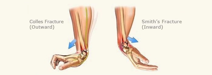
Distal Radius Fracture – Flexion Type
Much less common than the Colles Fracture, the reverse Colles or Smith fracture is due to a fall on the hand with the palm upwards. The hand is then forced into full flexion (bend) by the weight of the body. The bone fragments are pushed downwards and towards the palm.
Carpal Bone Fractures
There are a large number of injuries, including fractures, dislocations and sprains, which can occur to the wrist bones.
- Scaphoid fracture. The scaphoid is the most frequently injured of the wrist bones at around 60-70% of all such injuries. This fracture is also easy to miss, as it doesn’t show up on x-ray in about 10-15% of cases.
- Lunate fracture. The lunate is in the centre of the wrist bones and moves against the radius. A fall on the hand with the wrist right back or a fall on the heel of the hand can be the cause.
- Triquetrum fracture. This bone is quite commonly injured forced hyperextension (bending right back) of the wrist and movement inwards towards the ulnar direction. The local ligaments can be ruptured with a chip fracture of the bone.
- Capitate fracture. The capitate is the largest wrist bone. A direct blow or crushing force on the back of the hand or a fall on the outstretched hand can result in this injury.
- Hamate fracture. The hook of the hamate, a protruding part of the bone towards the palm side, is most commonly injured. Typical causes are hitting an object while holding a racquet, bat or club. Gripping causes pain in this injury.
- Trapezium fracture. Not common and occurs when the thumb is forced against the nearby wrist bone when it’s close to the palm.
- Trapezoid fracture. This is a rare fracture and can occur when a force is applied along the line of the second metacarpal, the bone that joins further up the hand with the index finger.
- Pisiform fracture. The pisiform is a small bone, which sits inside the tendon of a forearm muscle. This kind of bone is known as a sesamoid bone. A fall on the little finger side of the palm may cause a pisiform fracture.
- Dislocations. A large number of injury patterns can occur and often involve the lunate bone. Difficult to diagnose, it is important to get an expert opinion and the right management so that the wrist works normally again.
Treatment of Wrist Fracture
As with all suspected fractures, medical attention should be sought as soon as possible. X-ray will help confirm the nature of the injury and manipulation, plaster cast, surgery or a combination of treatments may be required.
After the injury has healed sufficiently, the cast or brace will be removed. Physiotherapy may be useful to regain any limited range of motion and to restore strength and normal function after a broken wrist.
Physiotherapy after Wrist Fracture
The need for physiotherapy after coming out of plaster varies widely. Some people, especially children, need no encouragement to regain hand function and may have little or no loss of wrist ranges of movement. Others may have significant problems and need intensive therapy.
Physiotherapy after a wrist fracture:
- The physio will check the ranges of movement of the neck, shoulder and elbow. The injury, followed by time in a cast or sling, may allow other joints to stiffen up. This occurs more often in the elderly.
- How the hand and wrist look is important. A “normal” result when the hand comes out of plaster is for it to look a bit scaly with dead skin, very dry and perhaps with some muscle wasting due to being in the cast. If the hand is very swollen and stiff, including the fingers, this will alert the physiotherapist to the possibility of complex regional pain syndrome (CRPS), a painful condition that needs early treatment.
- Some pain on movement is common, but it should not be severe and make the patient very anxious, otherwise the diagnosis of CRPS is again a question.
- The fingers should have a full or almost full range of movement. The wrist will likely be limited in flexion, extension and supination and pronation (turning the palm up and down when the arm is stuck out in front with the elbow bent).
- The hand and wrist should not be very sensitive to being touched and handled by the physiotherapist. If it’s very sensitive, then CRPS should again be considered.
- The physiotherapist in the fracture clinic will give advice on normal, two-handed, use of the arm, exercises and perhaps a soft splint for resting the hand for a few days. If the hand in very stiff or painful then referral for further physiotherapy is indicated.
References:
- Distal Radius Fractures – Broken Wrist. OrthoInfo. http://orthoinfo.aaos.org/topic.cfm?topic=a00412
- Wrist Fractures and Dislocations. Medscape. http://emedicine.medscape.com/article/1285825-overview#a5
Last Review Date: 19-02-2020
Next Review Date: 14-02-2022